
The Complete Ussing Chamber Guide

1) What an Ussing Chamber Actually Measures & Why
Goal. Quantify transepithelial transport and barrier integrity across a tissue or monolayer separating two solutions (mucosal/apical vs serosal/basolateral).
Primary electrical readouts
-
PD (Potential Difference, mV): open-circuit voltage across the epithelium.
-
Isc (Short-Circuit Current, µA/cm²): net active ion transport when PD is clamped to 0 mV.
-
TER / TEER (Ω, Ω·cm²): barrier tightness; higher values = tighter junctions.
-
TEER is area-normalized:
TEER = (R_sample − R_blank) × Area.
-
Flux readouts (chemical)
-
Paracellular tracer flux (e.g., mannitol, FITC-dextran, inulin): permeability of tight junctions.
-
Transcellular flux (e.g., glucose, amino acids): transporter-mediated uptake.
-
Apparent permeability (Papp) for a solute:
Papp = (dQ/dt) / (A × C0)
wheredQ/dt= appearance rate on receiver side,A= area,C0= donor concentration.
Why this matters. These metrics underpin research in CFTR/ENaC physiology, IBD, celiac disease, diarrhea models, airway hydration, skin barrier, and drug absorption/toxicity.
2) The Physics: Making Sense of PD, Isc, and TER
-
Ohm’s Law (tissue):
V = I × R.-
When voltage-clamped to 0 mV, the injected Isc equals net electrogenic transport (e.g., CFTR-mediated Cl⁻ secretion minus ENaC Na⁺ absorption).
-
-
Series resistances (solution + electrodes + bridges) sit outside the tissue.
-
Good chamber design uses voltage-sensing electrodes close to the tissue to minimize series drop.
-
-
AC vs DC TER.
-
DC pulse/step: inject a small current briefly, compute ΔV/ΔI.
-
AC impedance: small sinusoid (e.g., 1 kHz) can isolate epithelial resistance and capacitance if the electronics support it.
-
Corrections you should apply
-
Liquid junction potentials (LJP): arise at solution interfaces; reduce by matching ionic strength and using KCl agar bridges.
-
Offset correction: short the sensing electrodes in symmetric solutions; set baseline PD ≈ 0 mV before mounting tissue.
-
Area normalization: always report µA/cm² (Isc) and Ω·cm² (TEER) using the true aperture area.
3) Hardware: What Each Component Does
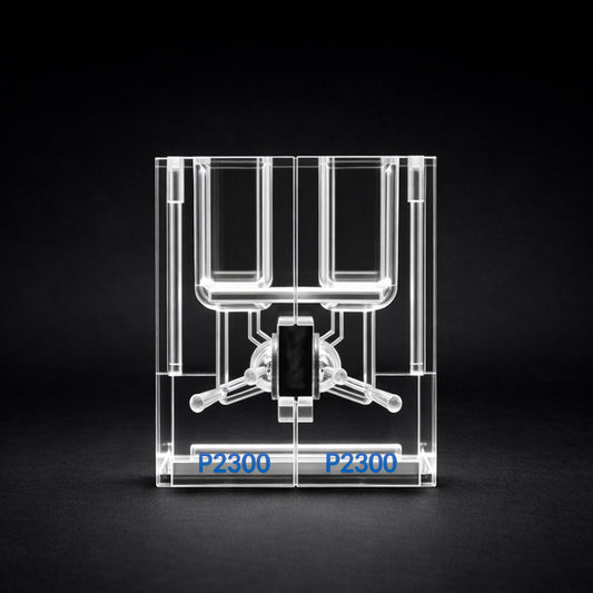
Chambers & Sliders (Apertures)
-
Classic split-halves = maximal flexibility; more handling skill required.
-
EasyMount = guided, fast, repeatable mounting; reduces tissue damage and variability.
-
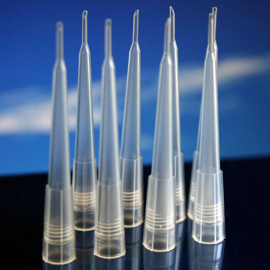
Aperture determines sensitivity and normalization (common: 0.071–0.64 cm²).
-
Gaskets (thickness, compliance) control compression; over-tightening causes edge leaks or ischemia.
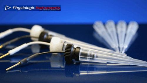
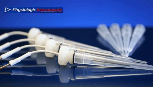
Electrodes & Bridges
-
Ag/AgCl electrodes via agar–KCl bridges (e.g., 3 M KCl in 2–3% agar).
-
Keep sensing electrodes close to tissue; current electrodes further out.
-
Maintenance: re-chloride Ag wires periodically; replace cloudy or dehydrated bridges.
Perfusion, Gas, Temperature
-
Gas lift with 95% O₂ / 5% CO₂ (carbogen) for bicarbonate buffers; gentle bubbling reduces unstirred layers.
-
Perfusion (constant or interval) prevents depletion/accumulation, keeps drugs at set concentrations.
-
Temperature: 37 °C with in-block or in-line heaters and calibrated probes; even 1–2 °C drift changes Isc/TEER.
Clamps & DAQ
-
Voltage–current clamps (single/multi-channel) measure PD, inject current, calculate Isc/TER automatically.
-
Data acquisition: time-stamped events, protocol steps, and annotations are critical for reproducibility.
Physiologic Instruments provides:
-
EasyMount and Classic Ussing Chambers (aperture options; tissue-specific sliders).
-
VCC series clamps (e.g., VCC MC8 multi-channel) for simultaneous experiments with integrated Isc/TER routines.
-
Perfusion manifolds, temperature controllers, gas lift assemblies, and Ag/AgCl electrode kits matched to the chambers.
4) Solutions & Recipes (working examples)
Verify with your IACUC/IRB/SOP. Buffer chemistry affects LJPs, transport driving forces, and viability.
Krebs–Ringer Bicarbonate (typical)
-
NaCl 117 mM, KCl 4.7 mM, CaCl₂ 2.5 mM, MgSO₄ 1.2 mM, KH₂PO₄ 1.2 mM, NaHCO₃ 25 mM, Glucose 10 mM
-
Gas with 95% O₂ / 5% CO₂, 37 °C.
Mannitol/Glucose Osmotic Pair (for symmetric osmolality)
-
Serosal: glucose 10 mM; Mucosal: mannitol 10 mM (or vice versa) to isolate transport asymmetries.
Common modulators
-
Amiloride (apical) to block ENaC (↓ Na⁺ absorption).
-
Forskolin/IBMX (stimulate cAMP → ↑ CFTR-mediated Cl⁻ secretion).
-
Bumetanide (basolateral) to block NKCC1 (limits Cl⁻ loading → ↓ secretion).
-
CFTRinh-172 to inhibit CFTR currents.
Tracer examples for flux
-
Paracellular: mannitol, FITC-dextran (3–4 kDa).
-
Transcellular: radiolabeled glucose, amino acids (observe safety protocols).
5) Protocol: Step-by-step SOP (field-tested)
A. Pre-run checks (10–20 min)
-
Electrode offset: immerse both sensing tips in the same buffer; adjust PD ≈ 0 mV.
-
Bridge health: gentle suction → free flow; no bubbles.
-
Temperature: stabilize chambers at 37 °C (±0.2 °C).
-
Solutions: pre-warm and gas; verify pH (7.3–7.4), osmolality (±5 mOsm).
-
Blank TER: assemble with a gasket/no tissue to measure system resistance if your SOP calls for R_blank.
B. Tissue/Insert prep (varies by model)
-
Native tissue: dissect carefully; avoid mesenteric tears; open along mesenteric border; trim to aperture.
-
Cell monolayers (e.g., Transwell): rinse, equilibrate 15–30 min in recording buffer before mounting (use adapter sliders if needed).
C. Mounting (2–5 min with EasyMount; longer with Classic)
-
Align tissue (mucosa → apical, serosa → basolateral).
-
Gently compress to seal—no wrinkles or edge extrusion.
-
Start perfusion/gas; wait 10–20 min for baseline stabilization.
D. Baseline acquisition
-
Record PD, Isc at steady state (drift <1%/min).
-
Measure TER via your clamp’s ΔI or AC routine; compute TEER.
E. Interventions (examples)
-
Amiloride (apical) → observe ↓ Isc (blocks ENaC).
-
Forskolin/IBMX (bilateral or basolateral) → ↑ Isc (activates CFTR).
-
Bumetanide (basolateral) → ↓ secretory current (blocks NKCC1).
-
Add test compound apical/serosal; track PD/Isc over time; collect samples for flux.
F. Flux sampling (if applicable)
-
Define intervals (e.g., every 10–15 min); maintain sink conditions (receiver side <10% of donor concentration).
-
Calculate Papp and clearance; normalize to area and time.
G. Close-out & QC
-
Recheck offset and blank to confirm system stability.
-
Log any temperature or pH deviations; inspect tissue for edge damage.
6) Data Analysis, Normalization & Reporting
Normalization
-
Isc to µA/cm²: divide by aperture area.
-
TEER to Ω·cm²:
(R_sample − R_blank) × Area. -
Report means ± SEM, n animals and n tissues per animal; avoid pseudo-replication (don’t treat multiple tissues from one animal as independent unless justified).
Typical plots
-
Time course of Isc with annotated additions.
-
TEER trajectory pre- and post-treatment.
-
Flux (cumulative or rate) vs time; bar charts of Papp.
Stats
-
Paired analysis when the same tissue serves as its own control.
-
Mixed models for multi-animal, multi-tissue designs.
-
Pre-define exclusion criteria (leaks, unstable baselines).
7) Troubleshooting (fast diagnostics)
| Symptom | Likely Cause | Fix |
|---|---|---|
| Large PD drift | Electrode offset, LJP changes, temp drift | Re-zero electrodes in symmetric buffer; verify 37 °C; match buffer composition |
| Noisy Isc | Bubbles at tissue, poor ground, mechanical vibration | Degas solutions; remove bubbles; secure cables; isolate pump vibration |
| Low/unstable TEER | Edge leak, over-compression, tissue damage | Remount with fresh gasket, reduce clamp force, trim cleaner edge |
| Isc saturates / can’t clamp | Bridge blockage, current electrode polarization | Replace bridges; clean current electrodes; check clamp compliance |
| Zero response to agonists | Tissue not viable, wrong side addition, expired reagents | Confirm viability markers; verify apical/serosal side; prepare fresh drugs |
8) Experimental Design by Tissue Type
Intestine/Colon
-
Pre-incubate with indomethacin (blocks prostanoids) if your model is prostaglandin-sensitive.
-
Gentle mucosal removal of contents; avoid epithelial tears.
Airway (trachea/bronchus)
-
ENaC block with amiloride (apical); CFTR activation via forskolin/IBMX; observe bumetanide sensitivity.
Skin
-
Thicker, higher TEER; ensure robust sealing; consider longer equilibration.
Cultured Monolayers (e.g., Caco-2, primary airway)
-
Verify confluence (pre-TEER thresholds); use adapter sliders; mind filter support resistance (subtract blank).
9) Quality & Reproducibility Checklist
-
Chamber identity, aperture, gasket lot recorded
-
Temperature/pH logs within range
-
Electrode offset before/after
-
Baseline stability (<1%/min drift)
-
Correct side additions and timestamps
-
Area normalization documented
-
Exclusion criteria predefined and applied
-
Raw files archived (time series + event log)
10) Safety & Compliance
-
Radiotracers: licensing, shielding, wipe tests, segregated waste.
-
Human/animal tissues: IRB/IACUC approvals, BSL-2 precautions as required.
-
Chemical hazards: bumetanide, forskolin, DMSO controls, proper SDS access.
11) Selecting & Budgeting the Right System
Match to goals
-
Teaching / pilot work: single channel, manual clamp, basic perfusion.
-
Core lab / pharma: multi-channel (4–8+), automated routines, integrated DAQ.
-
Barrier screening: stable TEER measurement with rapid mounting (EasyMount), reliable perfusion & temp control.
Cost expectations (configuration-dependent)
-
Entry single-channel: lower five figures
-
Standard multi-channel: mid–upper five figures
-
High-throughput automated: low six figures
Physiologic Instruments can specify:
-
EasyMount Ussing Chambers (fast, repeatable mounting for tissue and monolayers).
-
Classic Chambers (maximum flexibility for diverse tissues).
-
VCC MC8 (multi-channel clamp) and VCC 600 (single/dual) with built-in Isc/TER routines.
-
Perfusion manifolds, in-line heaters, gas lift kits, electrode/bridge kits, and software tuned for epithelial physiology.
Ask for an itemized quote matched to your tissue, aperture, and throughput.
12) Appendices
A) Quick-start SOP (one-page)
-
Warm/gas buffers; set 37 °C.
-
Zero electrodes in symmetric buffer.
-
Mount tissue (no wrinkles, correct orientation).
-
Stabilize; record baseline PD/Isc; measure TER → TEER.
-
Add modulators (amiloride → forskolin/IBMX → bumetanide, etc.).
-
Collect flux samples on schedule; keep sink conditions.
-
Close-out: re-check offsets; document exclusions.
B) Common Calculations
-
Isc density:
Isc_density (µA/cm²) = Isc_raw (µA) / Area (cm²) -
TEER:
TEER (Ω·cm²) = (R_sample − R_blank) × Area -
Papp:
Papp = (ΔQ/Δt) / (A × C0) -
Conductance (Gt):
Gt = 1 / R(normalize to area as needed)
C) Reporting Template (copy/paste)
-
Tissue: species/region; monolayer line & passage
-
Chamber: model, aperture (cm²), gasket thickness
-
Solutions: compositions, pH, temp; gas mix
-
Clamp: model, sampling rate, TER method (DC/AC)
-
Baseline PD/Isc/TEER ± SEM (n animals, n tissues/animal)
-
Agonists/antagonists: side, concentration, vehicle control
-
Flux: tracer, sampling schedule, Papp calculation
-
Statistics: tests, α level, exclusion criteria
13) Why Researchers Choose Physiologic Instruments
-
Purpose-built for epithelial physiology: chambers, clamps, and accessories designed to work together.
-
Reproducibility by design: EasyMount reduces handling variance; VCC clamps automate key steps (Isc/TER routines, annotations).
-
Scientist-to-scientist support: protocol tuning, aperture selection, and troubleshooting help from people who run these experiments.
-
Longevity & service: parts, electrodes, bridges, and upgrades available to keep systems in spec for years.
Ready to spec or standardize your lab’s setup? Physiologic Instruments can provide an itemized configuration tailored to your tissue, aperture, throughput, and analysis method—including training recommendations and a validation checklist.