

When Stable TEER Still Means Failing Tissue

Stable TEER does not always mean healthy
A stable TEER value does not guarantee a healthy or experimentally valid tissue preparation. TEER reflects electrical resistance stability, not full biological viability. A preparation can look calm on the trace while already being functionally compromised, which is why TEER should always be interpreted alongside functional response and tissue handling quality.
Key takeaways
- Stable TEER is not proof of tissue viability.
- Damaged tissue can still produce a flat TEER trace.
- Functional response matters as much as electrical stability.
- Handling, oxygenation, temperature, and mounting quality affect interpretation.
- TEER should be one checkpoint, not the entire conclusion.
One of the easiest mistakes to make in epithelial transport work is to treat a stable TEER reading as proof that the preparation is healthy. It feels reasonable. The value is not drifting, the trace looks calm, and nothing appears obviously wrong. For a principal investigator or a newer lab member, that kind of stability can look like experimental reassurance.
But TEER stability only tells you that resistance is not changing much over the time window you are observing. It does not, by itself, prove that the tissue is viable, responsive, well mounted, metabolically intact, or physiologically meaningful.
An epithelium can produce a steady number while already being compromised. The tissue may be ischemic, mechanically damaged, poorly oxygenated, mounted under tension, partially devitalized, or simply no longer capable of mounting a meaningful transport response. In those cases, the TEER value may remain flat while the biology underneath it is already failing.
This distinction matters because experimental decisions are often made too early. A stable baseline gets interpreted as a green light, and the rest of the protocol is built on a false assumption. By the time the problem becomes obvious, the experiment is already expensive, time consuming, and difficult to rescue.
What TEER actually tells you, and what it does not
TEER is useful because it provides a convenient readout of barrier resistance. It can help you compare preparations, monitor changes over time, and flag gross disruption of epithelial integrity. But it is only one layer of interpretation. It is not a complete measure of tissue quality.
| What TEER can help indicate | What TEER cannot prove on its own |
|---|---|
| Whether resistance is relatively high or low | Whether the tissue is metabolically healthy |
| Whether barrier properties changed during the run | Whether transport pathways are still functional |
| Whether gross leak developed | Whether the epithelium will respond normally to stimulation |
| Whether two conditions differ in apparent resistance | Whether the mounting, oxygenation, or handling were acceptable |
| Whether a preparation remained electrically stable | Whether the preparation remained biologically meaningful |
The core point is simple: electrical stability is not the same thing as physiological validity.
Why failing tissue can still look stable
There are several reasons a compromised preparation can still hold a relatively flat TEER value.
1. The tissue has stopped changing because it has already deteriorated
A dying or severely stressed epithelium does not always become noisy. Sometimes it becomes quiet. Once active processes are lost, the system may settle into a deceptively steady state. That state can be stable, but it is not healthy.
2. TEER reflects only one dimension of the preparation
A barrier can still show measurable resistance while secretory function, ion transport responsiveness, cellular energy state, or tissue architecture are already impaired. The number remains, but the experiment no longer represents normal physiology.
3. Mechanical problems may not produce immediate drift
Tissue that was stretched, nicked, compressed, or unevenly mounted can still produce a smooth baseline. The injury may not show up as immediate instability. Instead, it may reduce response amplitude, distort permeability interpretation, or create regional nonuniformity that TEER alone does not reveal.
4. Poor environmental support can create a false plateau
Inadequate oxygenation, temperature mismatch, or delayed processing can suppress biology without necessarily causing dramatic resistance movement. The tissue is underperforming, but the trace still looks calm.
The common false conclusion
The most common error is this: “The TEER was stable, so the tissue was fine.”
That conclusion collapses two different questions into one.
- Was the electrical measurement stable?
- Was the tissue biologically sound?
Those questions are related, but they are not identical.
Common interpretation mistakes
- Assuming a flat TEER trace automatically means the tissue is viable.
- Ignoring weak or absent functional response because the baseline looks stable.
- Overweighting baseline resistance and underweighting handling quality.
- Confusing grossly intact appearance with intact physiology.
- Failing to document collection delays, temperature drift, or poor oxygenation.
Signs that stable TEER may be misleading you
If a preparation shows a steady TEER value but any of the observations below are present, the tissue may still be compromised.
| Observation | Why it matters | Interpretation risk |
|---|---|---|
| Weak or absent response to expected stimulus | Suggests loss of functional transport capacity | You may wrongly conclude the biology is negative |
| Unusually low current changes despite normal-looking resistance | Can reflect damaged or exhausted epithelium | You may treat tissue failure as treatment failure |
| Delayed tissue processing before mounting | Increases ischemic stress and loss of viability | Stable TEER may hide pre-analytical damage |
| Mounting difficulty or visible edge stress | Can create nonuniform tissue behavior | Average resistance may look acceptable while local injury is severe |
| Poor oxygenation or temperature control | Suppresses active physiology | Flat TEER may be mistaken for healthy equilibrium |
| Tissue looks grossly intact but performs poorly | Appearance is not equivalent to function | Visual reassurance can override functional warning signs |
Quick troubleshooting guide
| Observation | Likely cause | What to check next |
|---|---|---|
| Stable TEER with weak agonist response | Loss of viability or suppressed transport function | Review handling time, oxygenation, temperature, and positive control response |
| Stable TEER with low current change | Functionally exhausted or injured tissue | Compare to expected response range and confirm tissue source quality |
| Stable TEER after difficult mounting | Mechanical compression or edge damage | Inspect mounting symmetry, edge seal, and tissue stress points |
| Stable TEER after processing delay | Ischemic compromise before chamber setup | Audit collection-to-mount timeline and holding conditions |
| Stable TEER with inconsistent downstream data | Preparation looks electrically calm but is biologically unstable | Check procedural consistency across tissues, chambers, and solutions |
A better framework for investigators
For a principal investigator building a lab culture, the goal is not to dismiss TEER. The goal is to place it in the right hierarchy. TEER should be treated as one checkpoint inside a broader tissue-quality framework.
A better rule is this: a stable TEER may be necessary in some experiments, but it is rarely sufficient evidence of tissue validity by itself.
Every preparation should be interpreted through at least three lenses.
| Lens | Question to ask | Example of useful check |
|---|---|---|
| Electrical | Is the reading stable and technically believable? | Baseline consistency, absence of obvious noise or drift |
| Functional | Does the tissue respond the way viable tissue should? | Expected current or transport response to a known perturbation |
| Procedural | Was the sample handled in a way that preserves biology? | Timing, oxygenation, temperature, mounting quality, solution quality |
When all three align, confidence increases. When TEER looks good but the other two do not, trust the discrepancy. Do not explain it away too quickly.
Healthy versus compromised tissue with stable TEER
| Feature | Healthy tissue with stable TEER | Compromised tissue with stable TEER |
|---|---|---|
| Response to agonist or known stimulus | Falls within expected range | Weak, delayed, or absent |
| Current behavior | Consistent with model and preparation | Blunted, inconsistent, or unexpectedly flat |
| Handling history | Prompt, controlled, and reproducible | Delayed, inconsistent, or stressful |
| Mounting quality | Smooth, symmetric, minimal edge stress | Difficult seal, compression, stretch, or visible stress |
| Confidence in interpretation | Higher because electrical and biological evidence align | Lower because stability conflicts with functional or procedural warning signs |
Examples of stable TEER with bad biology
Scenario 1: Ischemic delay before mounting
A tissue is collected, then sits too long before being mounted. By the time the run starts, the resistance value appears calm and does not drift much. The team feels encouraged. But the tissue shows blunted transport responses throughout the experiment. The stable TEER did not confirm viability. It only reflected a steady electrical state after physiological quality had already been lost.
Scenario 2: Mechanical handling damage
An epithelium is slightly compressed during mounting. The preparation seals and produces a plausible resistance value. Nothing looks chaotic. Later, permeability or secretory interpretation becomes confusing because the tissue is no longer behaving uniformly. Again, the calm baseline was real, but it was not enough.
Scenario 3: Environmental under-support
The chamber reaches a technically stable recording condition, but oxygenation or temperature support is suboptimal. The tissue is no longer operating near its best physiological state. TEER remains flat, yet responsiveness weakens. A clean trace can mask a weak preparation.
When stable TEER is actually reassuring
Stable TEER becomes more reassuring when it appears alongside good procedural control and expected biological performance. A calm trace carries more weight when the tissue was mounted promptly, oxygenation and temperature were controlled, the preparation responded normally to a known stimulus, and the data fit the expected physiology of the model. In other words, stable TEER is most useful when it confirms other evidence rather than replacing it.
Questions to ask before trusting a stable TEER value
Before accepting stable TEER as evidence that an experiment is on solid ground, review the preparation more critically.
- Was the tissue processed promptly and consistently?
- Was mounting smooth, symmetric, and free of obvious edge stress?
- Were temperature and oxygenation truly controlled, not just assumed?
- Does the preparation show expected responsiveness, not just baseline stability?
- Does the tissue behavior make biological sense in the context of the model?
- Would you still trust this preparation if TEER were the only reassuring signal?
That last question is especially useful because it forces interpretation beyond a single stable number.
Before you trust the data
- Tissue collected and mounted promptly
- Temperature confirmed throughout the experiment
- Oxygenation or gassing confirmed
- Mounting edges visually checked
- Expected control or stimulus response verified
- Baseline stable and technically believable
Suggested minimum reporting items
If the data will be used for training, internal review, or publication, record the minimum contextual details needed to interpret stable TEER correctly.
- Tissue type and source
- Time from collection to mounting
- Temperature conditions
- Oxygenation or gassing conditions
- Solution composition
- Baseline TEER
- Functional response check used
- Any exclusion criteria or handling deviations
How this mistake affects interpretation and publication quality
Misreading stable TEER has consequences beyond a single failed run. A lab may conclude that a treatment had no effect, that a model is inconsistent, or that a protocol is robust when the real issue was compromised tissue quality being mistaken for baseline validity.
That error propagates. It affects replication, student training, troubleshooting decisions, and manuscript quality. Principal investigators are especially vulnerable to this because they are often building systems, training personnel, and making protocol decisions at the same time. A stable trace can become psychologically attractive because it appears to reduce uncertainty. In reality, it may only hide it.
What to teach your lab
A strong lab culture does not teach people to chase stable numbers. It teaches them to ask whether those stable numbers still represent living, responsive, interpretable biology.
Instead of saying, “The TEER was steady, so we were fine,” the better internal language is: “The TEER was steady, now we need to determine whether the tissue was still functionally valid.”
That shift improves experimental judgment.
Final perspective
A steady TEER trace can indicate technical stability while the biology is already failing.
Stable TEER can be a helpful sign, but it is never the whole argument. Tissues fail quietly all the time. Some fail with noise and drift. Others fail with calm, convincing stability. The second case is often more dangerous because it creates false confidence.
The practical lesson is simple: do not confuse a steady barrier signal with a healthy preparation. Treat TEER as one readout within a broader biological assessment, and your interpretations will be more reliable, more reproducible, and harder to fool.
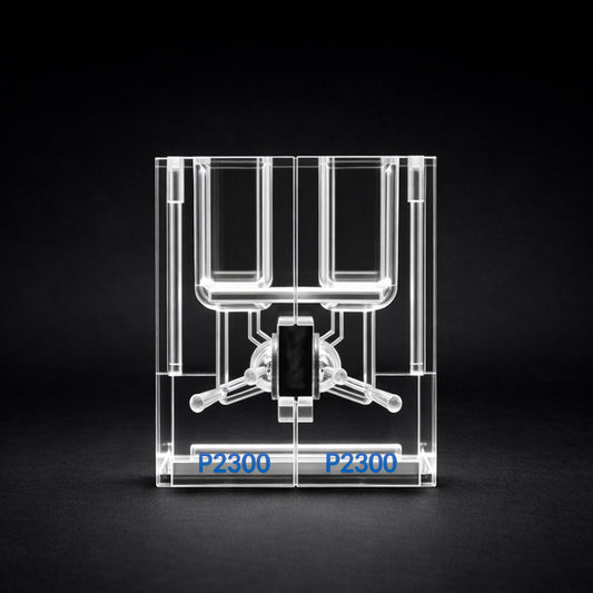
Conceptual illustration showing that a stable TEER signal can coexist with compromised epithelial biology and reduced functional performance.
Frequently Asked Questions
Does a stable TEER value always mean the tissue is healthy?
No. A stable TEER value only shows that resistance is not changing much during the time being observed. It does not prove that the tissue is viable, responsive, or still physiologically meaningful.
Can damaged tissue still produce a stable TEER reading?
Yes. Compromised tissue can still produce a flat, steady TEER value, especially if active transport processes have already declined or the preparation has settled into a nonresponsive state.
What does TEER measure well?
TEER is useful for assessing barrier resistance, comparing preparations, and tracking changes in epithelial integrity over time. It is a helpful electrical readout, but it is not a complete measure of tissue quality.
What can TEER not tell me by itself?
TEER alone cannot confirm metabolic health, proper mounting, adequate oxygenation, preserved transport function, or whether the tissue will respond normally to stimulation.
Why can failing tissue look electrically stable?
Failing tissue may appear stable because the preparation is no longer actively changing. Loss of responsiveness, mechanical damage, delayed mounting, or poor environmental support can all produce a deceptively calm trace.
What is the biggest interpretation mistake researchers make with TEER?
The most common mistake is assuming that a stable TEER reading means the tissue is fine. Electrical stability and biological validity are related, but they are not the same thing.
What warning signs suggest stable TEER may be misleading?
Weak responses to expected stimuli, unusually low current changes, delayed tissue processing, visible mounting stress, poor oxygenation, and grossly intact tissue that performs poorly are all warning signs.
Should TEER be used alone to judge tissue quality?
No. TEER should be interpreted alongside functional responses and procedural quality checks such as handling time, oxygenation, temperature control, and mounting quality.
How should a principal investigator evaluate tissue quality more reliably?
A stronger approach is to review the preparation through three lenses: electrical stability, functional responsiveness, and procedural quality. Confidence is much higher when all three align.
What is the practical takeaway for TEER experiments?
Do not confuse a steady TEER trace with a healthy preparation. Treat TEER as one readout within a broader biological assessment so the experiment is harder to misinterpret.