P2320A EasyMount Ussing Chamber Slider
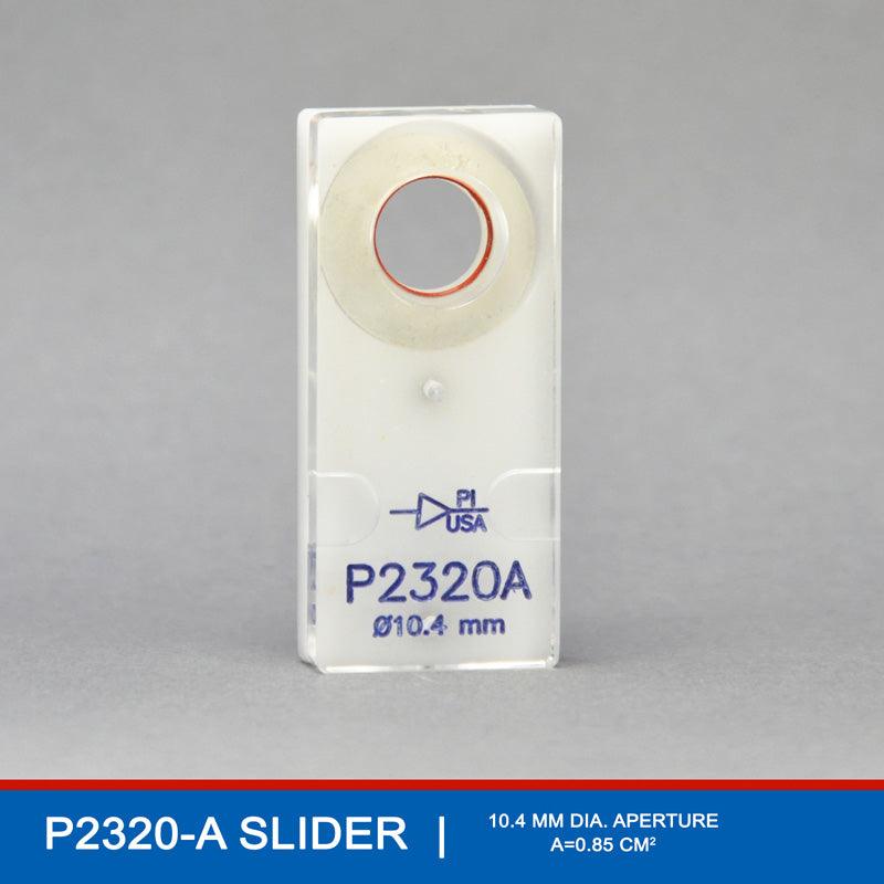
The P2320A EasyMount Ussing Chamber Slider is designed with a 10.4 mm diameter and an innovative curved mounting surface, making it ideal for studies involving human cornea and corneal endothelium. This slider’s unique curvature supports the natural shape of corneal tissues, minimizing edge stress and preserving tissue integrity, which is essential for accurate physiological measurements. With a 0.85 cm² aperture area, the P2320A allows precise study of transport, hydration, and permeability properties across corneal tissue. Its compatibility with the P2300 Ussing Chamber and specialized design make it a highly effective tool for ophthalmological research, enabling reliable, reproducible data collection in corneal barrier and transport studies.
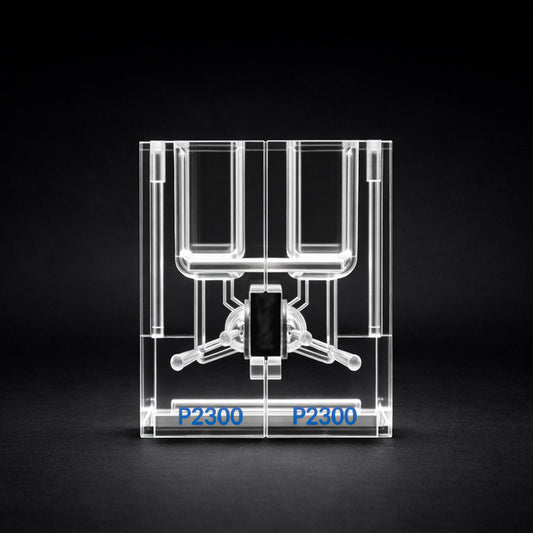
⦿ To be used with P2300 EasyMount Ussing Chambers- Regular price
- $353.00
- Regular price
-
- Sale price
- $353.00
- Unit price
- per
EasyMount Slider Details

P2320A EasyMount Ussing Chamber Slider for Studying Human Cornea & Corneal Endothelium
APETURE SIZE: 10.4MM | Area = 0.85 cm2
The P2320A EasyMount Ussing Chamber Slider is a precision-engineered 10.4 mm diameter tissue holder specifically designed for use with the EasyMount Ussing Chamber model P2300, tailored for studies involving the human cornea and corneal endothelium. With an aperture area of 0.85 cm², this slider offers an optimized surface for research into corneal transport and barrier properties. A unique feature of the P2320A slider is its curved mounting surface, which is particularly advantageous for securing and positioning the natural curvature of corneal tissue, thereby reducing stress on the tissue and preserving its physiological structure.
This curved design minimizes the risk of tissue distortion or edge compression, maintaining the cornea's structural integrity throughout the experiment and ensuring accurate measurement of transport and permeability parameters. When paired with the P2300 Ussing Chamber, the P2320A slider provides an ideal platform for researchers conducting in-depth studies of corneal hydration, ion transport, and drug permeability under controlled experimental conditions. Its specialized form makes it an invaluable tool for ophthalmologic research, providing reproducible and high-quality data in studies focused on human corneal health and barrier function.
P2320A EasyMount Ussing Chamber Slider Instructions
The P2320A EasyMount Ussing Chamber Slider is specifically designed to facilitate studies on the human cornea and corneal endothelium, with its defining feature being a curved mounting surface that closely matches the natural shape of these tissues. Here’s a guide on using the P2320A slider for experiments with the P2300 Ussing Chamber, from mounting the corneal tissue to conducting the experiment.
1. Preparing the Corneal Tissue Sample
- Tissue Collection and Preparation: Obtain a corneal sample suitable for transport studies, ensuring it includes the corneal epithelium or endothelium depending on the focus of the research. The tissue should be handled with care to maintain its natural curvature and hydration.
- Trimming: Trim the tissue to a size slightly larger than the 0.85 cm² aperture on the slider to ensure full coverage and a secure fit within the mounting area.
2. Mounting the Cornea on the P2320A Ussing Chamber Slider’s Curved Surface
- Curved Mounting Surface: The P2320A slider’s curved mounting surface is designed to match the natural contour of the corneal tissue, allowing it to sit securely without flattening. This curvature minimizes tension at the edges, reducing the risk of tearing or deformation and preserving the tissue's physiological structure for accurate transport measurements.
- Adhesion to the Slider: Use an appropriate, non-reactive adhesive, such as a medical-grade silicone adhesive, applied in a thin, even layer around the perimeter of the curved mounting surface. Avoid excessive glue near the aperture to keep the tissue’s central area clear for accurate measurements.
- Positioning the Tissue: Carefully position the cornea over the curved surface, pressing gently along the edges to ensure secure adhesion. The tissue should lie smoothly on the curved surface without wrinkles or air pockets, conforming naturally to the slider’s shape.
3. Mounting the Slider in the P2300 Ussing Chamber
- Preparing the Ussing Chamber: Fill each compartment of the P2300 Ussing Chamber with the appropriate solution for corneal studies, typically a balanced salt solution (BSS) to maintain corneal hydration and ion balance. Adjust the temperature to physiological levels, if necessary, to mimic in vivo conditions.
- Inserting the Slider: Place the P2320A Ussing chamber slider, with the mounted corneal tissue, into the chamber, ensuring a tight seal around the edges. The curved mounting surface will help keep the tissue in a natural position, preventing flattening and ensuring that both sides of the tissue are exposed to their respective compartments.
- Setting Up Electrodes and Sensors: Place voltage and current electrodes within each chamber compartment according to the P2300’s setup. This configuration allows for measurements of electrical properties, ion transport, or other parameters across the corneal tissue.
4. Conducting the Experiment
- Baseline Measurements: Record baseline parameters such as transepithelial resistance (TER) or potential difference (PD) to establish the initial physiological state of the corneal tissue.
- Experimental Treatments: Introduce any experimental agents, such as ion channel blockers or osmotic agents, to one side of the chamber if studying specific transport or permeability properties. This could include testing corneal hydration or permeability to drugs.
- Data Collection: Continuously monitor and record relevant parameters, such as TER, short-circuit current (Isc), or osmotic water flow, to assess the effects on corneal barrier function or ion transport in response to the experimental conditions.
5. Completing the Experiment and Cleaning
- Data Analysis: After concluding the experiment, save and analyze the data to interpret corneal transport and barrier properties.
- Disassembly and Cleaning: Remove the P2320A slider from the chamber, carefully detach the corneal tissue, and dispose of it according to laboratory protocols. Clean the slider, chamber compartments, and electrodes with distilled water followed by a suitable cleaning solution to prepare the equipment for future use.
The P2320A’s curved mounting surface is particularly advantageous for maintaining the natural curvature and structural integrity of corneal tissues, which is essential for accurate measurements in barrier and transport studies. This design supports detailed, reproducible research on human corneal health and provides valuable insights into physiological responses and permeability dynamics across corneal tissue.

