

How Electrophysiology Unlocks Epithelial Transport Insights

- ⚡ Understanding Electrophysiology
- 🧬 Why It Matters in Epithelial Transport
- 🔋 The Epithelial Barrier as an Electrical System
- 🎛️ Voltage vs. Current Clamping
- ⚗️ Ussing Chambers Overview
- 🧰 Anatomy of a Complete Setup
- 📟 Role of the VCC MC8 Amplifier
- 📈 Acquire & Analyze Workflow
- 📏 Key Parameters (Isc, PD, Gt)
- 🧪 Case Study: Chloride Secretion
- 🛠️ Stabilizing Recordings
- 🌬️ Beyond Intestine: Airway & Renal
- 🚀 Innovations & Emerging Models
- 🧭 Building a Reliable Workflow
- 🔗 Bringing It All Together
- ❓ FAQ
⚡ Understanding Electrophysiology: The Science of Bioelectric Signals
Electrophysiology quantifies how living tissues generate and respond to electrical signals arising from ion gradients across membranes. Movement of ions through channels, transporters, and paracellular pathways produces measurable voltages and currents that reflect underlying transport mechanisms.
🧬 Why Electrophysiology Matters in Epithelial Transport
Epithelia regulate sodium, chloride, potassium, and water flux to maintain composition of luminal and interstitial fluids. Electrophysiological recordings capture transepithelial signals as channels (e.g., ENaC, CFTR) and transporters respond to agonists, inhibitors, and changes in ionic gradients.
Because signals are quantitative and time-resolved, researchers can link molecular perturbations to functional outcomes such as absorption, secretion, and barrier integrity.
↑ Top🔋 The Epithelial Barrier as an Electrical System
An intact epithelium behaves like a circuit: tight junctions contribute resistance, electrogenic pumps establish driving forces, and channels carry current. The resulting transepithelial potential difference (PD or Vte) and short-circuit current (Isc) are reliable readouts of transport.
🎛️ Voltage vs. Current Clamping
In voltage clamp, the amplifier holds the transepithelial voltage constant and records the current required to maintain that voltage—revealing net ionic current. In current clamp, a defined current is applied and voltage changes are recorded—useful for following spontaneous or induced PD dynamics.
| Mode | Controlled | Measured | Typical Use |
|---|---|---|---|
| Voltage clamp | PD (Vte) | Isc | Quantify net ion transport; compute conductance (Gt) with pulses |
| Current clamp | I (applied) | PD | Follow PD responses to stimuli; characterize tissue excitability |
⚗️ Ussing Chambers Overview
Ussing chambers mount a tissue or cultured monolayer between two half-chambers with independent mucosal and serosal perfusion. Electrodes measure PD and deliver clamping current while solutions, gases, and temperature are precisely controlled.
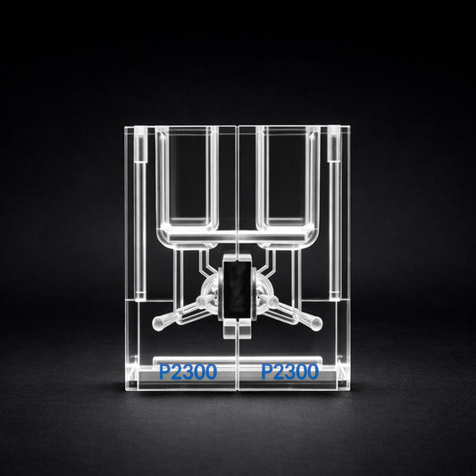
The EasyMount P2300 from Physiologic Instruments supports rapid mounting, reliable seals, and interchangeable sliders to match tissue geometry—facilitating reproducible readings across intestinal, airway, and renal epithelia.
↑ Top🧰 Anatomy of a Complete Setup
| Component | Purpose | Physiologic Instruments Example |
|---|---|---|
| Ussing chamber & sliders | Houses tissue; defines aperture; separates mucosal/serosal baths | EasyMount P2300 |
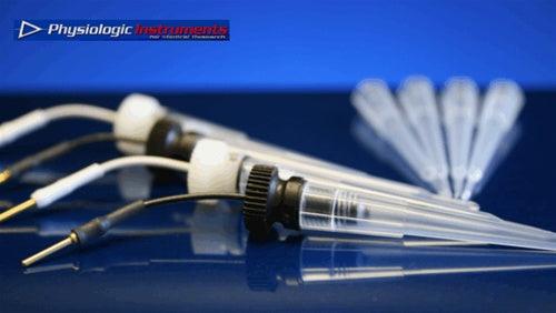
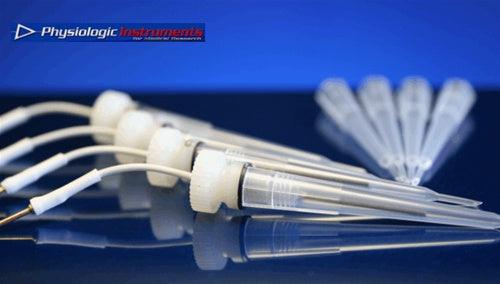
| Ag/AgCl voltage & current electrodes | Measure PD; pass current for clamp control | Matching electrode sets (PI catalog) |
| Clamp amplifier | Applies voltage/current clamp; computes Isc, Gt | VCC MC8 |
| Perfusion/heating & gas control | Maintains temperature, oxygenation, and composition | Chamber heater blocks & gas manifolds |
| Data acquisition & analysis | Record traces; automate calculations; export graphs | Acquire & Analyze |
📟 Role of the VCC MC8 Amplifier
The VCC MC8 provides voltage and current clamp control with multi-channel throughput for parallel experiments. Built-in pulse protocols enable conductance estimation, while low-noise front-ends help stabilize baseline Isc in challenging tissues.
| Capability | Benefit |
|---|---|
| Multi-channel operation | Higher throughput and internal replication |
| Programmable pulses | Rapid Gt assessment without manual switching |
| Clamp limit safeguards | Protects tissues from excessive current/voltage |
| Stable baseline control | Improves reproducibility of Isc measurements |
📈 Acquire & Analyze Workflow
Acquire & Analyze streamlines recording, annotation, and analysis. Typical steps: (1) establish baseline, (2) apply pulses for Gt, (3) add agonists/inhibitors, (4) annotate events, (5) average segments, (6) export figures and CSV.
📏 Key Parameters: Isc, PD, and Conductance (Gt)
| Parameter | What it indicates | Unit | Notes |
|---|---|---|---|
| Isc (short-circuit current) | Net active ion transport under PD = 0 | µA·cm−2 | Area-normalize to compare tissues/filters |
| PD (potential difference) | Transepithelial voltage at I = 0 | mV | Useful for tracking rapid responses |
| Gt (conductance) | Paracellular & transcellular pathway conductance | mS·cm−2 | Derived from voltage pulses; inversely related to Rt |
🧪 Case Study: Chloride Secretion in Intestinal Tissue
Chloride secretion can be probed by raising intracellular cAMP to activate CFTR and by isolating sodium currents with amiloride. A standard sequence is: amiloride (block ENaC) → forskolin/IBMX (activate CFTR) → CFTR inhibitor (confirm specificity).
| Step | Addition | Expected Isc/PD effect | Interpretation |
|---|---|---|---|
| 1 | Amiloride (apical) | Decrease Isc (blocks Na+ absorption) | Isolates secretory component |
| 2 | Forskolin ± IBMX | Increase Isc (stimulates CFTR-mediated Cl− secretion) | Demonstrates cAMP-driven secretion |
| 3 | CFTR inhibitor | Reduce Isc towards baseline | Confirms CFTR involvement |
🛠️ Stabilizing Recordings
| Issue | Likely cause | Action |
|---|---|---|
| Drifting baseline | Temperature fluctuation; slow electrode polarization | Allow thermal equilibration; precondition electrodes; verify ground |
| Noise spikes | Bubbles; perfusion turbulence; loose lead | Degas solutions; dampen flow; reseat connectors |
| Low signal | Leakage around mount; damaged tissue | Re-mount with correct slider; inspect seals; confirm viability |
| Clamp saturation | Excessive resistance or stimulus | Adjust limits on VCC MC8; reduce stimulus amplitude/duration |
🌬️ Beyond Intestine: Airway and Renal Models
Airway epithelia support coordinated Na+ absorption and Cl− secretion to regulate airway surface liquid, while renal epithelia enable fine control of electrolyte and water balance. Ussing chamber methods adapt to these tissues with appropriate solutions, gas mixtures, and sliders.
🚀 Innovations & Emerging Models
Advancements include organoid/3D culture interfaces, miniaturized sensors, and automated perfusion control. Analysis pipelines increasingly leverage event detection and templated workflows to streamline figure generation and data sharing.
🧭 Building a Reliable Workflow
- Prepare Define solutions, gas, and temperature; pre-check electrode offset.
- Mount Use the correct slider aperture and verify seals.
- Calibrate Zero PD; run brief pulses to estimate Gt.
- Record Stabilize baseline; document events in Acquire & Analyze.
- Validate Apply positive/negative controls to confirm pathway specificity.
- Report Normalize by area; include solution recipes, temperature, and pulse parameters.
🔗 Bringing It All Together
Electrophysiology connects epithelial structure to function through precise electrical measurements. With the EasyMount P2300, the VCC MC8, and Acquire & Analyze, investigators can mount tissues quickly, clamp confidently, and convert signals into clear conclusions.
❓ Frequently Asked Questions
What does Isc tell me about epithelial transport?
Under voltage clamp with PD = 0, Isc reflects net active ion transport. Changes in Isc after adding agonists or inhibitors indicate the direction and magnitude of pathway-specific flux.
When should I choose voltage clamp instead of current clamp?
Use voltage clamp to quantify transport (Isc) and compute conductance with pulses. Use current clamp to track PD responses when you prefer minimal imposed current.
How do I calculate conductance (Gt)?
Apply brief voltage steps around the holding level and measure the resulting current. Conductance is ΔI / ΔV, area-normalized to mS·cm−2.
What are common causes of unstable baselines?
Temperature drift, electrode polarization, bubbles, and leaks around the mount. Equilibrate temperature, precondition electrodes, degas solutions, and confirm seals.
Which Physiologic Instruments products fit a starter setup?
A typical configuration includes the EasyMount P2300, VCC MC8, matched Ag/AgCl electrodes, heating/gas control, and Acquire & Analyze for recording and analysis.
Recommended Equipment for This Research
| Equipment Category | Description | Link |
|---|---|---|
| Ussing Chamber Systems | Complete electrophysiology platforms for epithelial transport and barrier function studies. | Ussing Chamber Systems |
| Ussing Chambers (EasyMount & Classic) | Individual chambers for intestine, airway, renal, and custom tissues. | Ussing Chambers |
| Ussing Chamber Slider Inserts | Precision acrylic sliders for mounting tissues and replicating experimental geometries. | Ussing Chamber Slider Inserts |
| Voltage/Current Clamps (VCC MC8 Series) | Voltage clamp amplifiers for CFTR assays, TEER, and transepithelial measurements. | Voltage Clamps |
| Acquire & Analyze Software | Data acquisition and analysis software for epithelial electrophysiology experiments. | Acquire & Analyze |